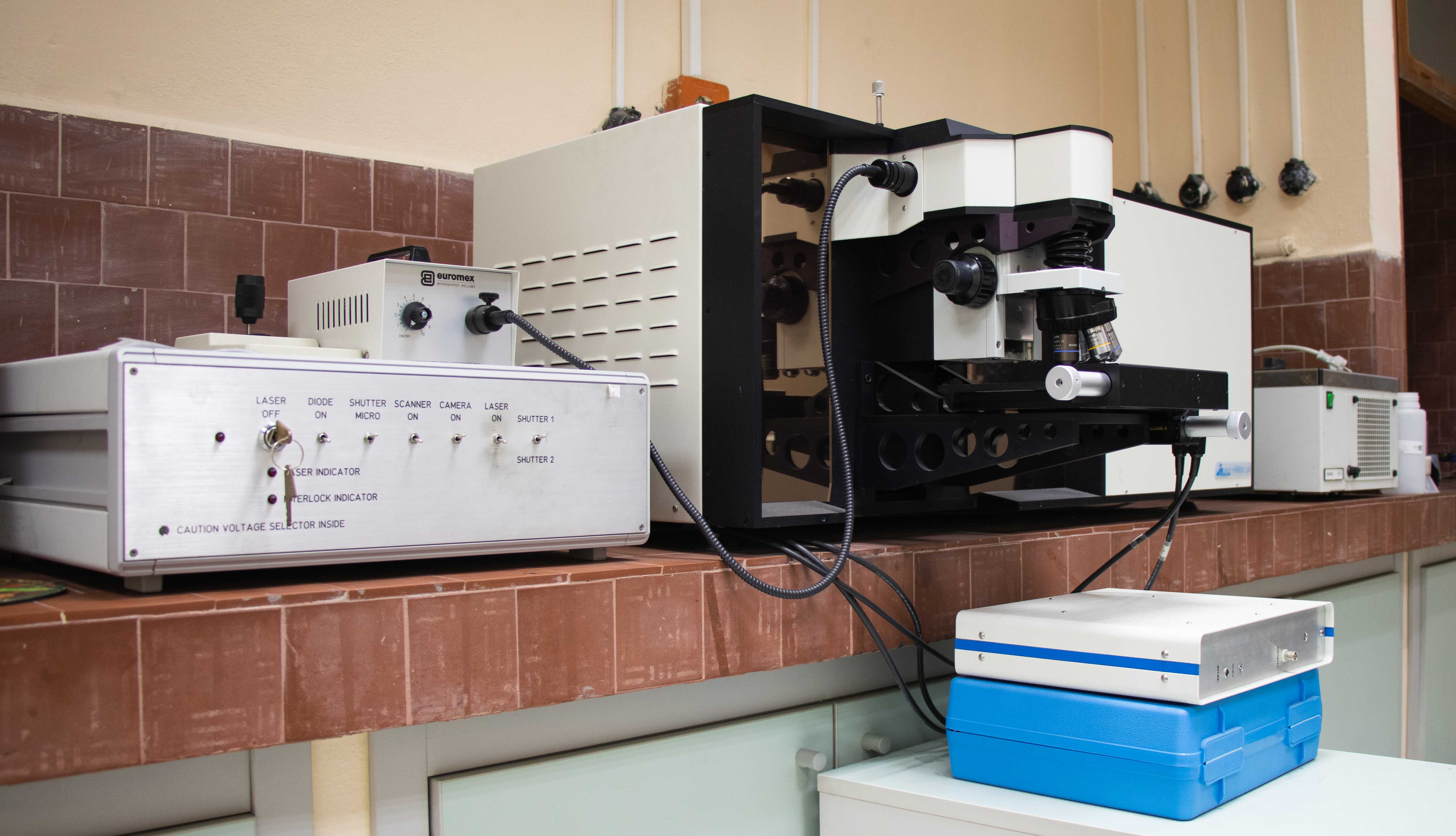
Raman spektrometresi, faz, kristalinite ve kimyasal yapı analizlerinde tahribatsız ölçüm sağlar.

|
Cihaz |
Raman Spektrometresi |
|
Marka |
HoribaJobin- Yvon HR800 |
Raman Spectroscopy is a non-destructive chemical analysis technique which provides detailed information about chemical structure, phase and polymorphy, crystallinity and molecular interactions. It is based upon the interaction of light with the chemical bonds within a material. Raman is a light scattering technique, whereby a molecule scatters incident light from a high intensity laser light source. Most of the scattered light is at the same wavelength as the laser source and does not provide useful information – this is called Rayleigh Scatter. However a small amount of lightis scattered at different wavelengths, which depend on the chemical structure of the analyte – this is called Raman Scatter. A Raman spectrum features a number of peaks, showing the intensity and wavelength position of the Raman scattered light. Each peak corresponds to a specific molecular bond vibration. In a Raman micro spectrometer the sample is illuminated with a laser beam through an objective of the integrated research grade optical microscope. Light from the focal spot is collected in back reflected mode with the same high quality microscope objective and sent through a spectrograph. Frequencies close to the laser line, due to elastic Rayleigh scattering, are filtered out while the rest of the collected light is dispersed onto a two dimensional detector (spectroscopic quality CCD).
| Ozellikler | |
| SpectralRange | 400 nm – near-Infrared (NIR) |
| Raman Shift | 100-6000 cm-1 |
| Spectral Resolution | depends on excitation frequency and grating used; Resolution = 0.16 cm-1at 633 nm excitation with 1800 gr/mm and CCD detector; Resolution = 0.30 cm-1at 785 nm excitation with 1800 ln/mm grating and CCD detector |
| Imaging Resolution | diffraction limited;702 nm using 633 nm with 50x N.A. 0.55 objective; 870 nm using 785 nm with 50x N.A. 0.55 objective |
| Detectors | JY open electrode CCD with enhanced quantum efficiency in the spectral range 450 – 950 nm; (2) InGaAs diode array JY IGA-3000, with highest sensitivity between 900 nm and 1700 nm |
İlgili Rehberler
Yüzey Karakterizasyon Yöntemleri
Görüntüleme, kimya ve topografyayı birleştiren genel yorumlama çerçevesine geçin.
SEM-EDS Nasıl Yorumlanır?
Kaplama ve bozunma davranışını okumak için morfoloji ile bileşimi birlikte değerlendirin.
XRD Nasıl Yorumlanır?
Kaplama yorumunu faz ve kristalografik bilgiyle destekleyin.
Profilometri Nedir?
Yükseklik haritalarından pürüzlülük yorumuna ve yüzey süreç kararlarına geçin.
XPS Nedir?
Yüzeye duyarlı kimyasal analizlerin oksit tabakaları ve arayüzleri nasıl çözdüğünü görün.